ICAP
ANA Patterns - Nuclear Patterns
Code Pattern Previous Nomenclature Description
AC-1
Nuclear homogeneous
Diffuse
Homogeneous and regular fluorescence across all nucleoplasm. The nucleoli maybe stained or not stained depending on cell substrate. Mitotic cells (metaphase, anaphase, and telophase) have the chromatin mass intensely stained in a homogeneous hyaline fashion.
AC-2
Nuclear dense fine speckled
none
Speckled pattern distributed throughout the interphase nucleus with characteristic heterogeneity in the size, brightness and distribution of the speckles. Throughout the interphase nucleus, there are some denser and looser areas of speckles (very characteristic feature). The metaphase plate depicts strong speckled pattern with some coarse speckles standing out.
AC-3
Centromere
kinetochore
Discrete coarse speckles (40-80/cell) scattered in interphase cells and aligned at the chromatin mass on mitotic cells. e.g. anti-CENP B
AC-4
Nuclear fine speckled
fine granular
Fine tiny speckles across all nucleoplasm. The nucleoli may be stained or not stained. Mitotic cells (metaphase, anaphase, and telophase) have the chromatin mass not stained. e.g. anti-SS-A/Ro, anti-SS-B/La
AC-5
Nuclear large/coarse speckled
spliceosome/nuclear matrix
Coarse speckles across all nucleoplasm. The nucleoli may be stained or not stained. Mitotic cells (metaphase, anaphase, and telophase) have the chromatin mass not stained. e.g. anti-Sm, anti-U1 RNP
AC-6
Multiple nuclear dots
6-20 nuclear dots, NSpI, PML bodies
Countable discrete nuclear speckles (6 to 20 nuclear dots/cell). e.g. SP-100
AC-7
Few nuclear dots
1 to 6 nuclear dots, coiled body, Cajal bodies
Countable discrete speckles (1 to 6 nuclear dots/cell in most cells). These are known as Cajal bodies or coiled bodies. e.g. anti-p80-coilin
AC-8
Homogeneous nucleolar
none
Diffuse fluorescence of the entire nucleolus, while the metaphase plate shows no staining. e.g. anti-PM-Scl, anti-Th/To.
AC-9
Clumpy nucleolar
none
Irregular staining of the nucleoli and Cajal bodies with a peri-chromosomal staining at the metaphase plates. e.g. anti-fibrillarin.
AC-10
Punctate nucleolar
nucleolar speckled
Densely distributed but distinct grains seen in the nucleoli of interphase cells. In metaphase cells, up to 5 bright pairs of the nucleolar organizer regions (NOR) can be seen within the chromatin body. The cytoplasm of mitotic cells may be slightly positive. e.g. anti-NOR-90, anti-RNA polymerase I
AC-11
Smooth nuclear envelope
nuclear rim, nuclear membrane, membranous
Homogeneous staining of the nucleus with greater intensity at its outer rim and no staining at the metaphase and anaphase chromatin plates. There is a peculiar accentuation of the fluorescence at the points where adjacent cells touch each other. e.g. anti-lamin B.
AC-12
Punctate nuclear envelope
nuclear membrane pores
Nuclear envelope reveals a punctate staining in interphase cells, with accentuation of fluorescence at the points where adjacent cells touch each other. No staining of the metaphase and anaphase chromatin plates. e.g. anti-gp210.
AC-13
PCNA-like
none
Pleomorphic speckled nucleoplasmic staining, with variability in size and brightness of the speckles. In interphase, some cells are negative (G1 phase), some are intensely stained (S-phase) and some present rare and scattered speckles with occasional nucleolar staining (late S and early G2 phases). Mitotic cells are not stained.
AC-14
CENP-F-like
MSA-3, NSp-II
Nuclear speckled pattern with striking variability in intensity with the strongest staining in G2 phase and weakest/negative staining in G1. The centromeres are positive only in prometaphase and metaphase, revealing multiple aligned small and faint dots. Prometaphase cells frequently show a weak staining of the nuclear envelope. During anaphase and telophase, some sera demonstrate intense staining in the ring located at the midzone (i.e. mid-body, stem body) where the division of the daughter cells is taking place. The surrounding cytoplasm of the mitotic cells is diffusely stained.
AC-29
Topo I-Like
Scl-70-like, Scl-86, DNA Topo I

The Topo I-like pattern can comprise staining of five subcellular regions:
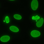
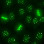
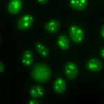
1) Prominent fine speckled nuclear staining in interphase cells.
2) Consistent strong fine speckled staining of condensed chromatin in mitotic cells. Depending on the serum dilution used, the mitotic chromatin staining may appear homogeneous.
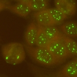
3) Strong staining of nucleolar organizing region (NOR).
4) Delicate and weak cytoplasmic weblike staining radiating from the perinuclear area to the vicinity of plasma membrane (in general, the cytoplasmic staining becomes more prominent during titering the sample to higher dilutions.
5) Inconsistent nucleolar staining that can appear as a punctate nucleolar or perinucleolar staining in interphase cells. Nucleolar staining is not a universal feature of this pattern.

See Andrade et al. for full discussion (Clin Chem Lab Med. 2018;56:1783-8.)


Online since 19 May 2015