ICAP
ANA Patterns - Search Result

Results for numa in Patterns

Code Pattern Previous Nomenclature Description  
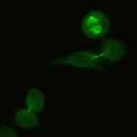
AC-25 Spindle fibers The spindle fibers between the poles are stained in mitotic cells, associated with cone-shaped decoration of the mitotic poles. Spindle fibers cover both NuMA-like and non-NuMA patterns. NuMA-like pattern has associated distinct nuclear speckles. Only non-NuMA patterns are shown here while NuMA-like patterns are shown in the NuMA-like group.
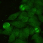
AC-26 NuMA-like MSA-1, centrophilin Nuclear speckled staining with spindle fibers.

Results for numa in FAQ's

Code Question Answer
18 Differences between multiple, mixed, and composite HEp-2 IFA patterns?
Question: Can you provide more information on how ICAP defines the differences between multiple patterns, mixed patterns, and composite patterns in HEp-2 IFA?

Answer: We refer to simple patterns when we observe single and isolated clear-cut HEp-2 IFA patterns, e.g. AC-23 or AC-3. The terms multiple, mixed, and composite IFA patterns refer to close but distinct concepts that go beyond the simple patterns.
1. The term "multiple patterns" applies to situations where two or more individual and clearly discernable patterns, e.g. AC-1 + AC-23 or AC-4 + AC-22, are observed in an individual serum sample (i.e., each of the patterns is clearly identified).
2. The term "mixed patterns" applies to observations where a serum sample produces more than one pattern in the same cell compartment and it is not possible to clearly identify each of the components. As an example, AC-4 + AC-5 can commonly be seen together but we are unable to distinguish each of the components (AC-4 and AC-5); in those cases, the HEp-2 IFA staining pattern is regarded as a mixed nuclear-speckled pattern.
3. Finally, the term "composite patterns" is applied to cases in which a single antibody causes staining of two or more cell compartments and the composite image is so characteristic that it always evokes the presence of that autoantibody. For example, the AC-26 pattern is an interphase nuclear staining pattern accompanied by mitotic spindle fiber staining and is typically associated with anti-NuMA antibodies. Similarly, the AC-14 pattern, typically associated with anti-CENP-F antibodies, is characterized by nuclear fine speckled staining with variable intensity in interphase cells, a delicate centromere staining only in mitotic cells, staining of the inter-cellular bridge, and nuclear envelope staining exclusively in prophase cells.


Online since 19 May 2015